
What are they epidermal nevus syndromes?
An epidermal nevus is a type of birthmark in which there is an overgrowth of one or more components of the epidermis or outer layer of the skin. Epidermal nevus syndromes refer to the coexistence of a keratinocytic epidermal nevus or an organoid nevus with other abnormalities in the skin and other organs. They are sometimes called systematized epidermals. naevi. They reflect mosaicism, in which there are two different cell lines with different genetics and they are due to the postzygotic. mutation.
Epidermal nevus syndromes generally arise sporadically, with the exception of CHILD syndrome, which family.
Several authors have commented that the term 'epidermal nevus syndrome' is outdated now that genetic The causes of many of the disorders are known.
Keratinocytic epidermal nevus syndromes
Keratinocytic epidermal nevus
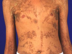
Systematized epidermal nevus
Three syndromes associated with a keratinocytic epidermal nevus are now well defined.
- BOYCongenital hemidysplasia with ichthyosiform nevus and lI am b reeffects) syndrome
- Type 2 segmental Cowden's disease
- Fibroblasts growth factor receiver three epidermal nevus syndrome
Child syndrome
CHILD syndrome is inherited as a dominant X-linked disorder and is fatal in males. CHILD means Congenital Hypoplasia with ichthyosiform nevus and limb defects. It is due to mutations at gene known as NSDHL (or NAD [P] steroid dehydrogenase H gene). This gene involved in metabolism of cholesterol.
The infantile nevus is variable, inflamed and covered by waxy yellowish scales. Usually strictly on one side of the body (most often the right) with a clear cut middle line demarcation, or you can follow the Blaschko lines or both. Often favors a body crease. the pathology shows characteristically sparkling histiocytes at dermal papillae. CHILD nevus can disappear spontaneously only to reappear later.
Other features may include:
- Abnormal thickening nail
- Strawberry-like lesions on the tips of the fingers
- Skeletal defects: shortening or absence of fingers or limbs
- Long bone x-ray changes - chondrodysplasia punctata
- The undergrowth of other bones
- Congenital defects of the heart, kidneys, or other organs.
- Neurological diseases
Segmental Cowden disease type 2
Multiple hamartoma Cowden syndrome or disease may include a Cowden nevus when called segmental Cowden disease type 2. Cowden naevus is a linearBushy, bumpy growth, like warts. PTEN Genetic mutations (phosphatase and tensin homolog), which are not present in Proteus syndrome, which can resemble segmental Cowden syndrome type 2 (see below), have been detected. the PTEN The normal function of the gene is to suppress tumors. Characteristics of type 2 segmental Cowden disease include:
- Linear Cowden's nevus
- Other nevi: vascular malformation, lymphatic malformation, lipomas
- Neurological defects including hydrocephalusseizures
- Limb and toe overgrowth
- Jejunal or colonic polyps.
Fibroblast growth factor receptor three epidermal nevus syndrome
Fibroblast growth factor receptor 3 (FGFR3) epidermal nevus syndrome (García-Hafner-Happle syndrome) is caused by an R248C mosaic mutation of FGFR3 Gene.
Organoid nevus syndromes
Sebaceous nevus
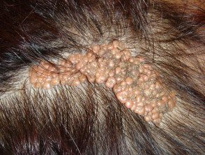
Sebaceous nevus
Schimmelpenning syndrome
Schimmelpenning syndrome has two Mendelian in Man online entries (OMIM 163200, 601359). Its other names include:
- Schimmelpenning-Feuerstein-Mims syndrome
- FMS syndrome
- Linear sebaceous nevus syndrome
- Jadassohn naevus fakomatosis
- Jadassohn's sebaceous naevus
- Organoid phakomatosis of the nevus.
Features include:
-
Sebaceous nevus, which may have minimal sebaceous overgrowth
- Skeletal defects: deformities of the face, trunk and extremities.
- Ocular Defects: conjunctival or corneal growth or coloboma
- Cerebral defects: mental deficiency, seizures, enlarged or shrunken brain tissue
- Hypophosphatemic rickets: vitamin D-deficient bone disease.
Phakomatosis pigmentokeratotica
Pigmentokeratotic phakomatosis (sometimes spelled phakomatosis) is an example of didimosis (twin spotting) because two distinct syndromes appear to overlap in one person. It is characterized by:
- Sebaceous nevus
- Papular naevus spilus (speckled lentiginous nevus).
Naevus spilus is a type of congenital melanocytic nevus. It comes as a flat coffee latte taint in a baby and then develops darker spots and papules.
Phakomatosis pigmentokeratotica can include features associated with Schimmelpenning syndrome (above) and papular nevus spilus syndrome. These are:
- Hyperhidrosis (excessive sweating)
- Muscular weakness
- Dysesthesia (sensory disturbance)
Many other abnormalities have been described in individual cases.
Didimosis aplasticosebacea
Didymosis aplasticosebacea is the association of sebaceous nevus with aplasia skin congenital
SCALP syndrome
SCALP syndrome is aplasticosebaceous didymosis in association with a giant melanocytic nevus. The name is an acronym for sebaceous nevus, central nervous system abnormalities, aplasia cutis, limbal dermoid, and Pigmented nevus.
Comedo nevus syndrome
The comedo naevus is composed of a group of open comedones plugged in with curb and it can arise on the head, trunk, or extremities. Comedo nevus has sometimes been reported in association with other defects:
- Waterfall on the same side as the nevus
- Bone defects on the same or opposite side.
- Girth of fingers and toes (syndactyly)
- Neurological defects on the same or opposite side.
Several other features have been described in individual cases.
Comedone naevus
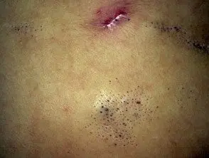
Comedo naevus
Angora hair nevus syndrome
The Angora capillary nevus is an epidermal nevus covered with long, smooth white hair. The angora hair nevus or Schauder syndrome may also include:
- Neurological defects, such as mental retardation, seizures, spasticity, hemiparesis.
- Optical defects, such as cataracts and coloboma.
- Skeletal defects: mainly facial malformations such as frontal protrusion, malformed ears and large tongue.
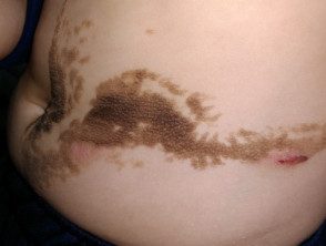
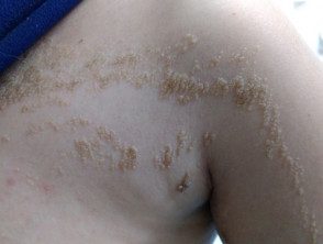
Becker naevus syndrome
The Becker naevus appears as patches of dark fur, often with a map or checkerboard outline. It is most commonly found on the upper back or shoulders. It tends to be more prominent in men due to the growth of dark hair at puberty. The syndrome can also include:
- Poor breast or nipple development on the affected side, more obvious in women.
- Supernumerary nipples
- waste of subcutaneous grease
- Axillary hair loss.
- Musculoskeletal abnormalities and asymmetry.
Becker naevus
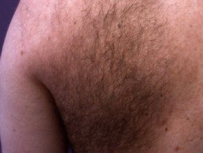
Becker naevus
Differential diagnosis epidermal nevus syndromes
Proteus syndrome
Proteus syndrome is characterized by excessive growth of the skin, connective tissue, fat, bones and other tissues.
- Flat, smooth and velvety keratinocytic epidermal nevus in 50% of cases
- Other nevi, eg vascular malformation, lymphatic malformation, skin aplasia, lipomas.
- Brain (cerebriform) overgrowth of palms or soles
- Enlarged fingers or toes
- Overgrowth of any other bone or tissue
- Neurological defects, including mild mental deficiencies and seizures
Other epidermal nevus syndromes
There are numerous individual case reports or small case series that do not fit the above descriptions. Some of these have been given names:
- Naevus trichilemmocysticus syndrome
- Gobello syndrome: epidermal nevus characterized by hypertrichosis and follicular hyperkeratosis in association with multiple bone defects.
- NEVADA Syndrome: Naevus Epidermicus Verrucosus with AngioDysplasia and Aneurysms
-
CLOVE syndrome: congenital lipomatous overgrowth, vascular malformations and epidermal nevus (similar to but different from Proteus syndrome).
Warty epidermal nevus with skeletal defects
Epidermal nevus syndrome
Epidermal nevus syndrome
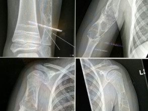
Epidermal nevus syndrome - skeletal defects
How are epidermal nevus syndromes diagnosed?
Epidermal nevus syndromes are diagnosed clinically by careful history and examination. Supportive investigations in a child with systemic or neurological symptoms may include:
- Skeletal survey
- Chest x-ray
- Computed tomography (Connecticut) scan or magnetic resonance image (Magnetic resonance)
- Molecular tests.
What is the treatment for epidermal nevus syndromes?
A multidisciplinary approach is often necessary to optimize symptom management. There is no cure.
